Technology is a gift to humanity. It has enabled our lives to become faster, smarter, and more efficient. Technology has optimized most of our everyday activities and made our lives easier. It has been of great benefit to the field of medicine. The advancement and changes have enabled more available treatment methods, safer surgeries, and even introduced new forms of drug delivery for best results. This advancement has increased our life expectancy and given hope for millions suffering from different ailments.
The field of dentistry is no exception to this technological evolution. There are various new treatment options available for treating common dental problems such as tooth sensitivity, decay, uneven tooth structure both for medical as well as cosmetic reasons. A bright smile can take you a long way in life. Today, with such unusual technological changes, we all can get a bright smile and look fabulous.
Dental scanners and X-ray
Just like how general x-rays assist doctors in understanding the problems inside your body, dental x-rays help to understand the root of the problem in your mouth. While specific issues appear on the layer, various serious problems are related to a bad tooth structure. Dental X-rays enable the doctor to view the growth and basic structure of your teeth and take the necessary action for treating the same. This type of X-rays become necessary before steps such as placing braces to make sure the alignment required is possible from this treatment.
Exploring Panoramic Dental Scanners
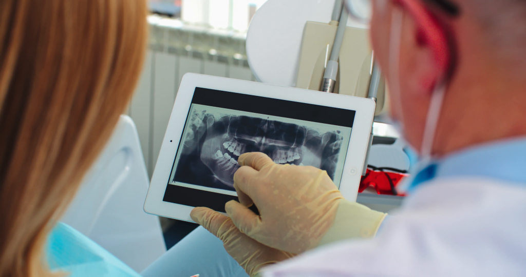
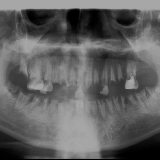
In the field of orthopantomography, panoramic radiography refers to a radiology technique. In this, a single scan can film the entire jawbones, teeth, mandible in one picture. It results in a 2D dental x-ray of the whole dental base. The process is non-invasive, and x-ray has been useful for acquiring an image of various parts of the body for many years now. This type of imaging gives a massive benefit to dentists who can get information for fitting implants, braces, prepare for root canal treatments. Apart from the diagnosis offered, this panoramic picture can give an excellent idea of any underlying problem that requires treatment.
The beauty of panoramic dental scanner lies in its ease of use. The panoramic scanner provides a flat image even of the curved structure like our jaw along with details of our bones and teeth. The advantage of Panoramic X-ray or scanners over traditional intraoral x-ray is that the imaging is from the outside. In the case of intraoral x-ray, the detector or film is inside the mouth, which could lead to discomfort.
Knowing about a Panoramic Dental Scanner
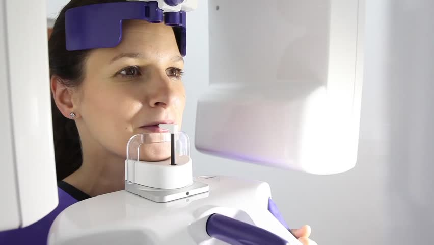
A panoramic dental x-ray or scan uses a small amount of ionizing radiation. The ionized particles from the scanner interact with the internal structure of the entire mouth and create one panoramic image. Dentists use these images for performing various procedures required as an action to treat specific problems. These scans happen on an everyday basis and require no preparation at all. The radiation levels are kept within safety considerations, and the patients usually use an apron made of lead to cover the rest of the body to prevent exposure. Simple precautions such as removing spectacles or jewellery or any other metal objects are essential to reduce risks. Although considered safe during pregnancy, it is advisable to stay clear of x-rays or scans during this period.
Today a dental panoramic scanner has advanced algorithms, which help provide best panoramic images of your dental structure even with minimum exposure. Few settings for capturing only the problem areas are also present in the modern system, which ensures reduced exposure and higher safety. These are light guided systems exclusive to the industry that aims at providing safety as well as better imaging quality in the predefined areas.
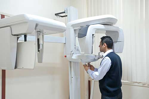
How does the Panoramic dental scanner look?
A typical panoramic x-ray or scanner machine consists of two sides. The x-ray tube is present on one-sided whereas the detector or film is present in the other. The patients sit with a bite blocker to open the mouth properly for a complete image. Also, the patient’s chin, forehead, and sides rest during the imaging. The technician will strap in the patient and secure the head is a proper position to acquire proper exposure. Modern units also have adjustments to accommodate patients in a wheelchair. It’s also essential for kids dental care.
How does it work?
X rays fall in the higher end of the light spectrum, where the energy is slightly larger than that of visible light. This energy enables the particles or waves to pass through objects, including our body. When directed properly, it can help examine the continuity or problems in the internal part of our body. A typical x-ray machine directs sudden bursts of radiation through the body and captures an image of the same.
In case of a panoramic x-ray or scan, the x-ray tube moves across the semi-circular perimeter of the head to get an image from jaw to jaw. The detector of film rotates on the other side of the head, which captures the image. The files are electronic and can be useful for initial diagnosis of problems in the bone structure or other dental issues. Advanced algorithms enable improving the image quality and acquiring better details, which help dentists understand the disease or problem better and offer immediate treatment options.
The Scanning Process
The complete process of acquiring the scan is simple and painless, and the entire exposure time lasts somewhere between 20-30 seconds per scan. Staying still is the key to obtaining a proper image, especially while the rotating arm travels around the perimeter of your head. This method is easier than intraoral x-rays, where people often complain of gag reflex and discomfort with the detector placed in their mouth.
Important points to know
A Panoramic Dental Scan can be beneficial for the initial diagnosis of the problem. Its imaging technique is not capable of embracing small details, especially in the tissues and muscles of the mouth. The curvature of the mouth also hinders during the imaging sometimes, causing blur images in these areas. There is a more advanced method such as CT (Computed Tomography or MRI (Magnetic resonance imaging), which offers a clearer picture, especially near the curvatures of our mouth for deeper details.
Panoramic scans or X-rays are affordable, and the facilities are available in various dental clinics and labs. With new algorithms and imaging techniques, the panoramic dental scanner might give better detailing, as well as depth in the years to come. This advancement could be a beneficial addition to the world of dentistry, as it can improve the quality of both diagnosis and expand possible treatment options.